


You’re Just Minutes Away from having your own Professional Documentation developed by our Health & Safety Consultants in London
Save time, money and avoid potential legal issues with Health & Safety Consultants London bespoke online services.
Whatever type of health & safety service you are after, our quick, reliable and affordable online system will help you get the health and safety documentation you need.
Safety Expert from Safety Expert on Vimeo.
What’s more, all of our services are pay-as-you-go, so you know exactly what you’re paying for – bespoke, expertly crafted and dependable policies for your business.
Need a Health & Safety Policy Fast?
As your business grows it becomes harder to ensure that a consistent attitude towards health and safety is maintained across the board.
Your business is legally required to comply with health and safety regulations – however, we believe that doing so shouldn’t be too complicated or overly expensive.
That’s where Safety Expert’s Health Safety Consultants London come in.
As Simple As One, Two, Three…
Our online system can provide you with the comprehensive health and safety services and documentation you need, including:
- Bespoke H&S Policies
- Health Safety Competent Person Service
- Risk Assessments
- COSHH Assessments
- Accident Report Forms
- Safety Inspection Checklists
- Health Safety Consultants London & Nationwide
- And Much More
Simply fill out our short online form and the Safety Expert system will instantly create your bespoke health & safety policy – without the need for endless meetings and expensive consultations.
Not sure where to begin? See our how to video here first!
View a list of some popular type health and safety policies offered by SafetyExpert here.
Need It Done Yesterday?
At SafetyExpert, we like to say that it’s better safe than sorry. We know how important your business is, which is why we believe you should have total peace of mind, should something go wrong. Accidents happen – but with our professional health and safety policies and other documentation, you’ll know you have the tools to deal with them effectively.
So why wait? No one can protect your business and create the tools you need to meet your legal requirements as quickly as us. Our questionnaire takes minutes to complete and documents are instantly downloadable meaning you can get back to working on the things that matter to your business. But as our customers will tell you, our turnaround time isn’t the only thing which sets us apart…
A Cut Above the Rest
We don’t believe in boggling our customers with legal jargon, neither do we like wagging the finger at them for not having adequate health and safety controls in place to begin with. Most importantly though, we like our customers to rest assured that they’re getting value for money. That’s why we offer you:
- Access to easy to use templates and checklists in order to help you assess your own compliance.
- The ability to access and amend your easy to edit documentation should you require, plus a guarantee that you can keep your documentation down the line.
- Impartial and confidential advice and guidance if and when you require it.
Need a more hands on service? Let us come to you.
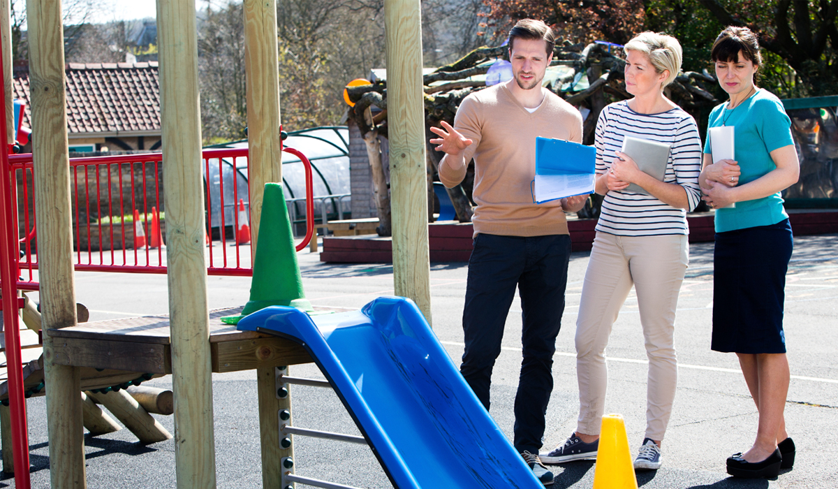
If you need a professional, fully-qualified health and safety consultant to visit you on-site then don’t hesitate to get in touch. Our consultants have long-standing industry expertise and provide a complete range of services – wherever you are in the United Kingdom as we don’t just have Health & Safety Consultants in London but nationwide!
Find out more about booking a Health & Safety Consultant visit.
Get In Touch – We Won’t Bite…
But if we did, you wouldn’t have to worry about needing an accident report form! To find out more, click here to visit our contact page – once you’ve filled in the details of your query, we’ll forward your details to the most suitable professional in our team and respond as quickly as possible…
At SafetyExpert, your safety is our concern – which is why we offer all of our customers a 100% satisfaction guarantee.
